Small animal magnetic resonance imaging (MRI):
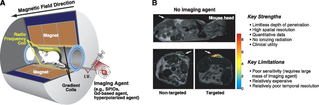
Figure A shows the basics of this technique. In general, an MRI scanner consists of a set of coils: a coil that produces a relatively homogeneous main magnetic field, gradient coils that produce variations in the magnetic field in the X, Y, and Z directions that It is used to locate the source of the MR signal, and finally the RF coils that generate an RF pulse that changes the state of the magnetic dipoles.
During an MRI scan, a living organism is placed inside a magnet (in this example, a mouse). Unpaired spins inside the body, known as magnetic dipoles, are aligned either parallel or in the opposite direction to the magnetic field. The MR signal is produced by a very small difference in the number of antiparallel and antiparallel spins (the vast majority of spins are parallel).
Figure B shows MRI images from a study to evaluate a new molecular MRI imaging agent in a mouse model of glioma. The image above is a cross-sectional image of a mouse, in which the tumor is marked with an arrow. The lower part of the coronal images shows the same mouse imaged 3 hours after injection of non-targeted (left) and chlorotoxin-targeting (glioma surface peptide) superparamagnetic iron oxide (SPIO) nanoparticles. Tumor image contrast enhancement is much greater for mice injected with targeted SPIO nanoparticles. These images demonstrate the advantage of using targeted MRI imaging agents to image biochemical targets in living organisms.
Ref: James, Michelle L., and Sanjiv S. Gambhir. A molecular imaging primer: modalities, imaging agents, and applications. Physiological reviews 92.2 (2012)
Persian Gulf Nuclear Medicine Research Center
Membership link: https://t.me/joinchat/AAAAAEC0ftOmHCJhRlKNyw
This post is written by Abbasifard_A